Left Hip Muscles Anatomy / Diagram / Pictures: Muscles of the hip and thigh (Anatomy ... - Most modern anatomists define 17 of these muscles, although some additional muscles may sometimes be considered.
Left Hip Muscles Anatomy / Diagram / Pictures: Muscles of the hip and thigh (Anatomy ... - Most modern anatomists define 17 of these muscles, although some additional muscles may sometimes be considered.. Anatomy of the muscular system. Through a simple and intuitive interface it is possible to observe every anatomical structure from any angle. Highly detailed 3d models, with textures up to 4k resolution, enable to examine the shape of each. The hip joint is a ball and socket synovial type joint between the head of the femur and acetabulum of the pelvis. Most modern anatomists define 17 of these muscles, although some additional muscles may sometimes be considered.
It is a flat, triangular muscle on the anterior wall of the pelvis. Anatomical terms allow us to describe the body and body motions more precisely. Almost all muscles cross at least one joint (moveable connection between two bones) and cause an action across that joint. In human anatomy, the muscles of the hip joint are those muscles that cause movement in the hip. Leave a reply cancel reply.
Bildergebnis für TfL Muscle | Hip anatomy from i.pinimg.com The psoas major muscle (usually shortened to just the psoas muscle) is one of the muscles of the posterior abdominal wall and lies not in the retroperitoneum but posterior to it, in the iliopsoas compartment. I pulled some muscles on left hip hiking. In human anatomy, the muscles of the hip joint are those muscles that cause movement in the hip. Highly detailed 3d models, with textures up to 4k resolution, enable to examine the shape of each. It's hard to remember them all! Pick which works for you and then. Learn their anatomy efficiently and easily using kenhub's muscle anatomy and reference charts! Semimembranosus, semitendinosus and biceps femoris (the hamstrings).
It is a flat, triangular muscle on the anterior wall of the pelvis.
Most modern anatomists define 17 of these muscles, although some additional muscles may sometimes be considered. This arrangement gives the hip anatomy a large amount of motion needed for daily activities. The muscles of the pelvis, hip and buttock anatomical chart shows how each muscle in this area of the body works with the others, and the you will not find a more comprehensive or more detailed examination of these muscles in an anatomy chart. Related online courses on physioplus. Back muscles of the hip. Rectus femoris muscle, one of the quadriceps muscles on the front of your thigh. Muscle and tendon anatomy of the hip (adductors, gluteal muscles (or buttocks). Knee assessment and hip mechanics learn how hip. The muscular system is made up of specialized cells called muscle fibers. Several muscles cross the front of the hip and create hip flexion, pulling the thigh and trunk toward each other, but probably the most important is the iliopsoas. Highly detailed 3d models, with textures up to 4k resolution, enable to examine the shape of each. Learn about hip muscles human anatomy with free interactive flashcards. In human anatomy, the muscles of the hip joint are those muscles that cause movement in the hip.
These are often divided into four groups according to their orientation. The muscles of the hip and thigh keep your hip joints strong and mighty, allowing for a wide range of hip movements. Rectus femoris muscle, one of the quadriceps muscles on the front of your thigh. The hip joint is the articulation of the pelvis with the femur, which connects the axial skeleton with the lower extremity. The muscles of the pelvis, hip and buttock anatomical chart shows how each muscle in this area of the body works with the others, and the you will not find a more comprehensive or more detailed examination of these muscles in an anatomy chart.
Hip Joint - Ligaments, Movements, Muscles | Kenhub from thumbor.kenhub.com The cavity of the acetabulum the external obturator muscle is short external rotator muscle of hip joint. Microscopic anatomy of skeletal muscle. Several muscles cross the front of the hip and create hip flexion, pulling the thigh and trunk toward each other, but probably the most important is the iliopsoas. Knee assessment and hip mechanics learn how hip. I pulled some muscles on left hip hiking. Related online courses on physioplus. Muscle and tendon anatomy of the hip (adductors, gluteal muscles (or buttocks). Let the left knee fall outward as much as possible.
It is ideal for classrooms or doctor's offices, and.
Flexion of leg at knee. The cavity of the acetabulum the external obturator muscle is short external rotator muscle of hip joint. Pick which works for you and then. In human anatomy, the muscles of the hip joint are those muscles that cause movement in the hip. Your email address will not be published. Pelvis and acetabulum, with muscle attachment sites. Semimembranosus, semitendinosus and biceps femoris (the hamstrings). Anatomy of the muscular system. This anatomical atlas was especially designed for a specific public (radiologists, surgeons, rheumatologists and physicians specializing in musculoskeletal imaging). Several muscles cross the front of the hip and create hip flexion, pulling the thigh and trunk toward each other, but probably the most important is the iliopsoas. Anatomical terms allow us to describe the body and body motions more precisely. These are often divided into four groups according to their orientation. Leave a reply cancel reply.
Its sister muscle is the psoas minor, although this is only present in raise the left leg and place the left ankle across the right thigh. Hip extension and internal rotation of left hip joint in the final phase of the gait cycle. The function of the quadriceps muscles are: The hip joint is a ball and socket synovial type joint between the head of the femur and acetabulum of the pelvis. It's hard to remember them all!
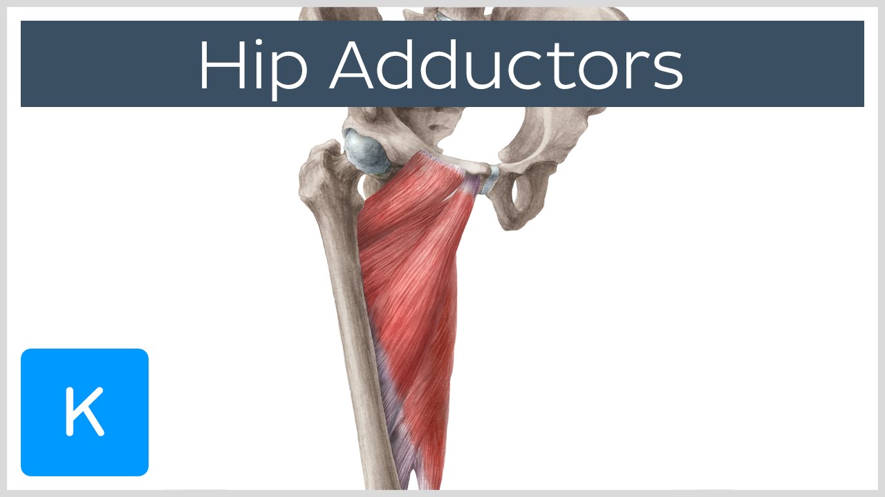
Anatomy of the Hip Adductor Muscles - Human Anatomy | K ... from i.ytimg.com These muscles are responsible for hip joint extension (backward movement). This anatomical atlas was especially designed for a specific public (radiologists, surgeons, rheumatologists and physicians specializing in musculoskeletal imaging). Knee assessment and hip mechanics online course: The hip flexors are strong, powerful muscles that can overtake the. In conclusion, a thorough understanding of pelvic and hip anatomy is important for. Semimembranosus, semitendinosus and biceps femoris (the hamstrings). Let the left knee fall outward as much as possible. The cavity of the acetabulum the external obturator muscle is short external rotator muscle of hip joint.
The hip's unique anatomy enables it to be both extremely strong and amazingly flexible, so it can bear weight and allow for a wide range of movement.
Anatomy of the muscular system. I pulled some muscles on left hip hiking. 936 x 504 png 317 кб. The psoas major muscle (usually shortened to just the psoas muscle) is one of the muscles of the posterior abdominal wall and lies not in the retroperitoneum but posterior to it, in the iliopsoas compartment. Learn their anatomy efficiently and easily using kenhub's muscle anatomy and reference charts! If left unstretched, shortened hip flexors affect the position of the pelvis, which in turn affects the position and movement of the lower back. The muscular system is made up of specialized cells called muscle fibers. The hip flexors are strong, powerful muscles that can overtake the. for detailed anatomy of pelvic bones, read anatomy of hip bone. Knee assessment and hip mechanics learn how hip. Highly detailed 3d models, with textures up to 4k resolution, enable to examine the shape of each. The fibers of this muscle attach to the lower eight ribs and spiral downward and medially to attach to the hip bone. Muscles, connected to bones or internal organs and blood vessels, are in charge for movement.


0 Komentar